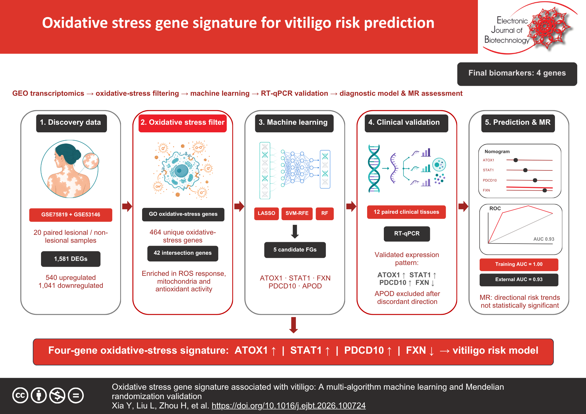
Graphical abstract
Keywords
Categories
How to Cite
Abstract
Background: Vitiligo is a common depigmentation disorder that affects approximately 1–2% of the global population. Oxidative stress plays a crucial role in its pathogenesis, yet systematic identification of oxidative stress-related biomarkers remains limited. This study aimed to identify and validate oxidative stress-related biomarkers for vitiligo and to construct a risk prediction model using bioinformatics, machine learning, and Mendelian randomization approaches.
Results: Through integration of high-throughput sequencing data, 1,581 differentially expressed genes were identified, of which 42 intersected with known oxidative stress-related genes. Three machine learning algorithms converged on five candidate genes. Real-time polymerase chain reaction validation in paired vitiligo lesional and non-lesional skin tissues confirmed the significant upregulation of ATOX1, STAT1, and PDCD10 and downregulation of FXN (p < 0.05). A risk prediction nomogram based on these four genes achieved high accuracy in the training dataset (area under the curve = 1.00) and an independent validation dataset (area under the curve = 0.93). PDCD10 demonstrated the strongest individual discriminatory performance (area under the curve = 0.820) in external validation. Mendelian randomization analysis indicated that ATOX1, STAT1, and PDCD10 had odds ratios greater than 1, suggesting a potential trend toward increased vitiligo risk, although the associations did not reach statistical significance.
Conclusions: This study identified ATOX1, STAT1, FXN, and PDCD10 as oxidative stress-related biomarkers for vitiligo. The four-gene risk prediction model demonstrated promising accuracy, with potential implications for early detection and personalized treatment strategies, pending further validation in larger cohorts.
References
AL-smadi K, Imran M, Leite-Silva VR, et al. Vitiligo: A review of aetiology, pathogenesis, treatment, and psychosocial impact. Cosmetics 2023;10(3):84. https://doi.org/10.3390/cosmetics10030084
Xu W, Qiu Z, Li C, et al. Recurrence and risk factors in cured patients with vitiligo: A real-life single-center retrospective study. Journal of Cosmetic Dermatology 2023;22(5):1680-1684. https://doi.org/10.1111/jocd.15637 PMid: 36721299
Spritz, RA, Santorico SA. The genetic basis of vitiligo. Journal of Investigative Dermatology 2021;141(2):265-273. https://doi.org/10.1016/j.jid.2020.06.004. PMid: 32778407
Roberts GHL, Santorico SA, Spritz RA. Deep genotype imputation captures virtually all heritability of autoimmune vitiligo. Human Molecular Genetics 2020;29(5):859-863. https://doi.org/10.1093/hmg/ddaa005 PMid: 31943001
Bergqvist C, Ezzedine K. Vitiligo: A focus on pathogenesis and its therapeutic implications. The Journal of Dermatology 2021;48(3):252-270. https://doi.org/10.1111/1346-8138.15743 PMid: 33404102
Li S, Zhu G, Yang Y, et al. Oxidative stress drives CD8+ T-cell skin trafficking in patients with vitiligo through CXCL16 upregulation by activating the unfolded protein response in keratinocytes. Journal of Allergy and Clinical Immunology 2017;140(1):177-189.e9. https://doi.org/10.1016/j.jaci.2016.10.013 PMid: 27826097
Zhang J, Hu W, Wang P, et al. Research progress on targeted antioxidant therapy and vitiligo. Oxidative Medicine and Cell Longevity 2022;2022:1821780. https://doi.org/10.1155/2022/1821780 PMid: 35320978
Chen J, Li S, Li C. Mechanisms of melanocyte death in vitiligo. Medicinal Research Reviews 2021;41(2):1138-1166. https://doi.org/10.1002/med.21754 PMid: 33200838
Xu X, Zhang C, Jiang M, et al. Impact of treatment delays on vitiligo during the COVID-19 pandemic: A retrospective study. Dermatologic Therapy 2021;34(4):e15014. https://doi.org/10.1111/dth.15014 PMid: 34075666
Mohammadi S, Amiri R, Khalili M, et al. Treatment protocols and efficacy of combined laser with medical treatment modalities in vitiligo. Journal of Cosmetic Dermatology 2022;21(8):3272-3291. https://doi.org/10.1111/jocd.14602 PMid: 34766697
Zhang J, Yu R, Guo X, et al. Identification of TYR, TYRP1, DCT and LARP7 as related biomarkers and immune infiltration characteristics of vitiligo via comprehensive strategies. Bioengineered 2021;12(1):2214-2227. https://doi.org/10.1080/21655979.2021.1933743 PMid: 34107850
Miao Y, Su D, Fu Q, et al. Identification of CKS2 and RRM2 as potential markers of vitiligo using bioinformatics analysis. Medicine 2022;101(46):e31908. https://doi.org/10.1097/MD.0000000000031908 PMid: 36401415
Li S, Dai W, Wang S, et al. Clinical significance of serum oxidative stress markers to assess disease activity and severity in patients with non-segmental vitiligo. Frontiers in Cell and Developmental Biology 2021;9:739413. https://doi.org/10.3389/fcell.2021.739413 PMid: 34977005
Atas H, Kocabiyik M, Gonul M, et al. Usefulness of ischemia-modified albumin in predicting oxidative stress in patients with vitiligo. Biomarkers in Medicine 2017;11(6):439-449. https://doi.org/10.2217/bmm-2016-0259 PMid: 28598198
Frisoli ML, Essien K, Harris JE. Vitiligo: Mechanisms of pathogenesis and treatment. Annual Review of Immunology 2020;38(1):621-648. https://doi.org/10.1146/annurev-immunol-100919-023531 PMid: 32017656
Guarneri F, Bertino L, Pioggia G, et al. Therapies with antioxidant potential in psoriasis, vitiligo, and lichen planus. Antioxidants 2021;10(7):1087. https://doi.org/10.3390/antiox10071087 PMid: 34356320
Karagaiah P, Valle Y, Sigova J, et al. Emerging drugs for the treatment of vitiligo. Expert Opinion on Emerging Drugs 2020;25(1):7-24. https://doi.org/10.1080/14728214.2020.1712358 PMid: 31958256
Lee JH, Kwon HS, Jung HM, et al. Treatment outcomes of topical calcineurin inhibitor therapy for patients with vitiligo: A systematic review and meta-analysis. JAMA Dermatology 2019;155(8):929-938. https://doi.org/10.1001/jamadermatol.2019.0696 PMid: 31141108
Chang HC, Hsu YP, Huang YC. The effectiveness of topical calcineurin inhibitors compared with topical corticosteroids in the treatment of vitiligo: A systematic review and meta-analysis. Journal of the American Academy of Dermatology 2020;82(1):243-245. https://doi.org/10.1016/j.jaad.2019.07.108 PMid: 31408687
Zhou Y, Khan M, Jiang L, et al. The current status of antioxidants in the treatment of vitiligo in China. Oxidative Medicine and Cell Longevity 2022;2022:2994558. https://doi.org/10.1155/2022/2994558 PMid: 35251468
Barrett T, Wilhite SE, Ledoux P, et al. NCBI GEO: Archive for functional genomics data sets—update. Nucleic Acids Research 2013;41(D1):D991-D995. https://doi.org/10.1093/nar/gks1193 PMid: 23193258
Leek JT, Johnson WE, Parker HS, et al. The sva package for removing batch effects and other unwanted variation in high-throughput experiments. Bioinformatics 2012;28(6):882-883. https://doi.org/10.1093/bioinformatics/bts034 PMid: 22257669
Ritchie ME, Phipson B, Wu D, et al. limma powers differential expression analyses for RNA-sequencing and microarray studies. Nucleic Acids Research 2015;43(7):e47. https://doi.org/10.1093/nar/gkv007 PMid: 25605792
Subramanian A, Tamayo P, Mootha VK, et al. Gene set enrichment analysis: A knowledge-based approach for interpreting genome-wide expression profiles. Proceedings of the National Academy of Sciences of the United States of America 2005;102(43):15545-15550. https://doi.org/10.1073/pnas.0506580102 PMid: 16199517
Ashburner M, Ball CA, Blake JA, et al. Gene Ontology: Tool for the unification of biology. Nature Genetics 2000;25(1):25-29. https://doi.org/10.1038/75556 PMid: 10802651
Kanehisa M, Goto S. KEGG: Kyoto Encyclopedia of Genes and Genomes. Nucleic Acids Research 2000;28(1):27-30. https://doi.org/10.1093/nar/28.1.27 PMid: 10592173
Yu G, Wang LG, Han Y, et al. clusterProfiler: An R Package for comparing biological themes among gene clusters. OMICS: A Journal of Integrative Biology 2012;16(5):284-287. https://doi.org/10.1089/omi.2011.0118 PMid: 22455463
Carbon S, Ireland A, Mungall CJ, et al. AmiGO: online access to ontology and annotation data. Bioinformatics 2009;25(2):288-289. https://doi.org/10.1093/bioinformatics/btn615 PMid: 19033274
Saeys Y, Inza I, Larrañaga P. A review of feature selection techniques in bioinformatics. Bioinformatics 2007;23(19):2507-2517. https://doi.org/10.1093/bioinformatics/btm344 PMid: 17720704
Parsad D, Gupta S. Standard guidelines of care for vitiligo surgery. Indian Journal of Dermatology, Venereology and Leprology 2008;74(7):37-45. PMid: 18688102
Lin Z, Qi R, Deng H, et al. Bidirectional mendelian randomization suggests causal effects of 731 immunophenotypes on vitiligo pathogenesis. Clinical, Cosmetic and Investigational Dermatology 2026;19:603361. https://doi.org/10.2147/CCID.S603361 PMid: 42145868
Hemani G, Zheng J, Elsworth B, et al. The MR-Base platform supports systematic causal inference across the human phenome. Elife 2018;7:e34408. https://doi.org/10.7554/eLife.34408 PMid: 29846171
Dell'Anna ML, Maresca V, Briganti S, et al. Mitochondrial impairment in peripheral blood mononuclear cells during the active phase of vitiligo. Journal of Investigative Dermatology 2001;117(4):908-913. https://doi.org/10.1046/j.0022-202x.2001.01459.x PMid: 11676831
Boniface K, Passeron T, Seneschal J, et al. Targeting innate immunity to combat cutaneous stress: The vitiligo perspective. Frontiers in Immunology 2021;12:613056. https://doi.org/10.3389/fimmu.2021.613056 PMid: 33936032
Khaitan BK, Sindhuja T. Autoimmunity in vitiligo: Therapeutic implications and opportunities. Autoimmunity Reviews 2022;21(1):102932. https://doi.org/10.1016/j.autrev.2021.102932 PMid: 34506987
Wang Y, Li S, Li C. Perspectives of new advances in the pathogenesis of vitiligo: From oxidative stress to autoimmunity. Medical Science Monitor 2019;25:1017-1023. https://doi.org/10.12659/MSM.914898 PMid: 30723188
Abdelsalam M, Allam SH, Zohdy M, et al. TLR4 gene polymorphisms in Egyptian vitiligo patients: Insights into emerging association with clinical activity, family history, and response to therapy. Journal of Genetic Engineering and Biotechnology 2021;19(1):132. https://doi.org/10.1186/s43141-021-00218-y PMid: 34468896
Yu N, Zhang S, Sun T, et al. Double-stranded RNA induces melanocyte death via activation of Toll-like receptor 3. Experimental Dermatology 2011;20(2):134139. https://doi.org/10.1111/j.1600-0625.2010.01208.x PMid: 21255093
Sun L, Sun J, Huo X, et al. Lipopolysaccharide reduces melanin synthesis in vitiligo melanocytes by regulating autophagy. Experimental Dermatology 2022;31(10):1579-1585. https://doi.org/10.1111/exd.14629 PMid: 35733278
Song PI, Abraham TA, Park Y, et al. The expression of functional LPS receptor proteins CD14 and toll-like receptor 4 in human corneal cells. Investigative Ophthalmology & Visual Science 2001;42(12):2867-2877. PMid: 11687531
Dong J, An X, Zhong H, et al. Interleukin-22 participates in the inflammatory process of vitiligo. Oncotarget 2017;8(65):109161-109174. https://doi.org/10.18632/oncotarget.22644 PMid: 29312598
Shi F, Kong BW, Song JJ, et al. Understanding mechanisms of vitiligo development in Smyth line of chickens by transcriptomic microarray analysis of evolving autoimmune lesions. BMC Immunology 2012;13:18. https://doi.org/10.1186/1471-2172-13-18 PMid: 22500953
Ungureanu L, Vasilovici AF, Halmagyi SR, et al. The many faces of autoimmune-mediated melanocyte destruction in melanoma. Frontiers in Immunology 2024;15:1417273. https://doi.org/10.3389/fimmu.2024.1417273 PMid: 39421737
Strassner JP, Harris JE. Understanding mechanisms of autoimmunity through translational research in vitiligo. Current Opinion in Immunology 2016;43:81-88. https://doi.org/10.1016/j.coi.2016.09.008 PMid: 27764715
Yang L, Wei Y, Sun Y, et al. Interferon-gamma inhibits melanogenesis and induces apoptosis in melanocytes: a pivotal role of CD8+ cytotoxic T lymphocytes in vitiligo. Acta Dermato-Venereologica 2015;95(6):664-670. https://doi.org/10.2340/00015555-2080 PMid: 25721262
Rashighi M, Agarwal P, Richmond JM, et al. CXCL10 is critical for the progression and maintenance of depigmentation in a mouse model of vitiligo. Science Translational Medicine 2014;6(223):223ra23. https://doi.org/10.1126/scitranslmed.3007811 PMid: 24523323
Wang XX, Wang QQ, Wu JQ, et al. Increased expression of CXCR3 and its ligands in patients with vitiligo and CXCL10 as a potential clinical marker for vitiligo. British Journal of Dermatology 2016;174(6):1318-1326. https://doi.org/10.1111/bjd.14416. PMid: 26801009
He S, Xu J, Wu J. The promising role of chemokines in vitiligo: From oxidative stress to the autoimmune response. Oxidative Medicine and Cellular Longevity 2022;2022:8796735. https://doi.org/10.1155/2022/8796735 PMid: 35096274
Xu P, Xue YN, Ji HH, et al. H2O2-induced oxidative stress disrupts mitochondrial functions and impairs migratory potential of human epidermal melanocytes. Experimental Dermatology 2020;29(8):733-741. https://doi.org/10.1111/exd.14134 PMid: 32580253
Wang Y, Li S, Li C. Clinical features, immunopathogenesis, and therapeutic strategies in vitiligo. Clinical Reviews in Allergy & Immunology 2021;61(3):299-323. https://doi.org/10.1007/s12016-021-08868-z PMid: 34283349
Page S, Chandhoke V, Baranova A. Melanin and melanogenesis in adipose tissue: Possible mechanisms for abating oxidative stress and inflammation? Obesity Reviews 2011;12(5):e21-e31. https://doi.org/10.1111/j.1467-789X.2010.00773.x PMid: 20576005
Tanacan E, Atakan N. Higher incidence of metabolic syndrome components in vitiligo patients: A prospective cross-sectional study. Anais Brasileiros de Dermatologia 2020;95(2):165-172. https://doi.org/10.1016/j.abd.2019.07.006 PMid: 32113676
Wu M, Wang L, Wu H, et al. Leptin deficiency in CD8+ T cells ameliorates non-segmental vitiligo by reducing interferon-gamma and Granzyme B. Frontiers in Immunology 2023;14:1158883. https://doi.org/10.3389/fimmu.2023.1158883 PMid: 37207234
Xu M, Shi J, Min Z, et al. A network pharmacology approach to uncover the molecular mechanisms of herbal formula Kang-bai-ling for treatment of vitiligo. Evidence-Based Complementary and Alternative Medicine 2019;2019:3053458. https://doi.org/10.1155/2019/3053458 PMid: 31781265
Vo TTT, Peng TY, Nguyen TH, et al. The crosstalk between copper-induced oxidative stress and cuproptosis: A novel potential anticancer paradigm. Cell Communication and Signaling 2024;22(1):353. https://doi.org/10.1186/s12964-024-01726-3 PMid: 38970072
Yang D, Xiao P, Qiu B, et al. Copper chaperone antioxidant 1: Multiple roles and a potential therapeutic target. Journal of Molecular Medicine 2023;101(5):527-542. https://doi.org/10.1007/s00109-023-02311-w PMid: 37017692
Sudhahar V, Shi Y, Kaplan JH, et al. Whole-transcriptome sequencing analyses of nuclear antixoxidant-1 in endothelial cells: Role in inflammation and atherosclerosis. Cells 2022;11(18):2919. https://doi.org/10.3390/cells11182919 PMid: 36139494
Jin W, Zhao J, Yang E, et al. Neuronal STAT3/HIF-1?/PTRF axis-mediated bioenergetic disturbance exacerbates cerebral ischemia-reperfusion injury via PLA2G4A. Theranostics 2022;12(7):3196-3216. https://doi.org/10.7150/thno.71029 PMid: 35547748
Butturini E, Carcereri de Prati A, Mariotto S. Redox regulation of STAT1 and STAT3 signaling. International Journal of Molecular Sciences 2020;21(19):7034. https://doi.org/10.3390/ijms21197034 PMid: 32987855
Ast T, Meisel JD, Patra S, et al. Hypoxia rescues frataxin loss by restoring iron sulfur cluster biogenesis. Cell 2019;177(6):1507-1521e16. https://doi.org/10.1016/j.cell.2019.03.045 PMid: 31031004
Moreno-Lorite J, Pérez-Luz S, Katsu-Jiménez Y, et al. DNA repair pathways are altered in neural cell models of frataxin deficiency. Molecular and Cellular Neuroscience 2021;111:103587. https://doi.org/10.1016/j.mcn.2020.103587 PMid: 33418083
Valentino M, E. Dejana E, Malinverno M. The multifaceted PDCD10/CCM3 gene. Genes & Diseases 2021;8(6):798-813. https://doi.org/10.1016/j.gendis.2020.12.008 PMid: 34522709
Bajo-Grañeras R, Sanchez D, Gutierrez G, et al. Apolipoprotein D alters the early transcriptional response to oxidative stress in the adult cerebellum. Journal of Neurochemistry 2011;117(6):949-960. https://doi.org/10.1111/j.1471-4159.2011.07266.x PMid: 21463325
Bajo-Grañeras R, Crespo-Sanjuan J, García-Centeno RM, et al. Expression and potential role of apolipoprotein D on the death-survival balance of human colorectal cancer cells under oxidative stress conditions. International Journal of Colorectal Disease 2013;28(6):751-766. https://doi.org/10.1007/s00384-012-1616-2 PMid: 23296401

This work is licensed under a Creative Commons Attribution-ShareAlike 4.0 International License.
Copyright (c) 2026 Electronic Journal of Biotechnology